what went wrong questions
What Does Yellow or Brown Discolouration Mean?
A peptide solid should be white to off-white. Distinct yellow or brown means a new chemical has formed, most often from oxidised tryptophan or from a reaction between amines and a reducing sugar. What each route implies, when colour is there by design, and why colour is a poor measure of how much has changed.
It means a new chemical has formed that absorbs visible light, because peptide solids themselves are white to off-white. In a peptide the two usual culprits are oxidation of tryptophan, whose breakdown products are yellow to brown, and a Maillard-type reaction between the peptide's amine groups and a reducing sugar where one is present in the formulation 13. A few materials are coloured by design, and for those colour means nothing. For everything else, distinct yellow or brown is a genuine sign of chemical change.
What colour does not tell you is how much has changed. A strongly coloured product can be visible at low levels, and the commonest forms of degradation produce no colour at all. So discolouration is a reason to stop and record, not a measurement, and its absence is not reassurance.
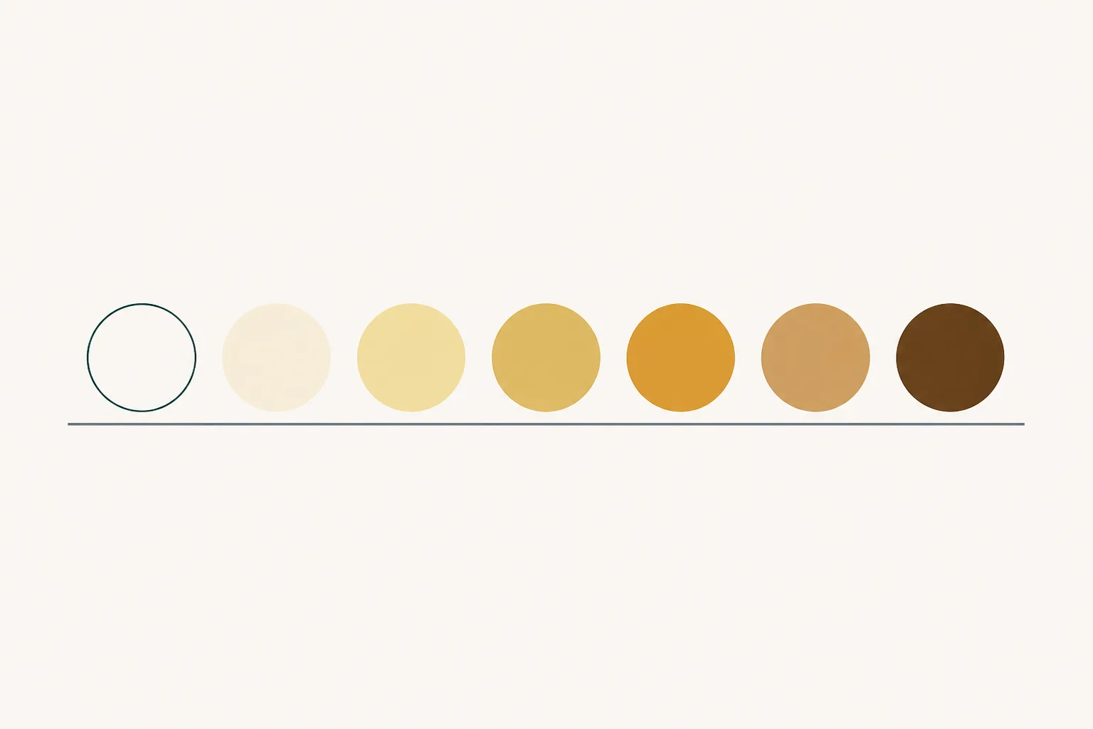
What colour should a peptide be?
White to off-white as a dry solid, and colourless in solution, for the great majority of peptides. The amino acids that make up peptides do not absorb visible light, and neither do the peptide bonds that join them, so there is nothing in an intact sequence to give it colour. The slight cream or ivory tone some batches show is within the ordinary range.
The right reference point is the batch's own documentation. Specifications for drug substances include a description of the material's appearance, its physical state and colour, as a basic test 5, and certificates of analysis for research peptides commonly carry an equivalent appearance line. If the certificate says white to off-white powder and the vial holds something plainly yellow or tan, the material no longer matches its own description.
Why does oxidation turn a peptide yellow?
Because oxidising tryptophan converts it into products that absorb light towards the visible range. Tryptophan's indole ring can be opened by oxidation to give N-formylkynurenine and kynurenine, among other products, and these absorb at longer wavelengths than tryptophan itself does 1. When enough of them accumulate, the material looks yellow and, as the reaction goes further, brown.
This is not speculation drawn from general chemistry. Analytical work on a therapeutic protein that had changed colour after exposure to light and heat identified tryptophan-derived oxidation products as the chromophores responsible, and located them at specific tryptophan residues in the molecule 1. The same chemistry applies to any peptide that contains tryptophan.
Oxidation of this kind is driven by light, by dissolved oxygen, by trace metal ions and by peroxide impurities, and it proceeds far more readily in solution than in a dry, sealed solid 2. That makes a yellowed solution a more familiar sight than a yellowed powder, and a yellowed powder a stronger signal that the material has had an unusual history, such as exposure to light, warmth, air or moisture.
Not all oxidation is visible. Methionine is the residue most readily oxidised, and its product, methionine sulfoxide, is colourless. Cysteine oxidation is colourless as well 2. A peptide containing methionine but no tryptophan can therefore be heavily oxidised without changing colour at all, which is one reason colour is a poor general-purpose indicator.
What is a Maillard reaction doing in a peptide vial?
Reacting a sugar with the peptide's free amine groups, if a reducing sugar is present. The Maillard reaction is the family of reactions between reducing sugars and amines, familiar as the browning of cooked food, and it happens in pharmaceutical formulations too. The amine groups most exposed to it are the peptide's free N-terminus and the side chain of lysine.
A study of a freeze-dried peptide hormone formulated with different sugars found rapid covalent modification of the molecule when glucose was the excipient, with glucose adducts forming on its amino groups through the Maillard reaction, and showed that the reaction proceeded in the dry, lyophilised state rather than requiring solution 3. The early adducts are colourless. Browning comes from the later stages, when the initial products rearrange and condense into coloured compounds, which is why discolouration by this route tends to indicate that the reaction has gone a long way.
Which sugar matters. Glucose, lactose and maltose are reducing sugars and can take part directly. Sucrose and trehalose are not reducing, and neither is mannitol, which is why they are favoured as bulking agents and stabilisers in freeze-dried products; sucrose can, however, hydrolyse into reducing sugars under acidic conditions 36. Most research peptides are supplied with no sugar at all, in which case this route is simply unavailable and oxidation is the more likely explanation for colour.
Moisture and warmth accelerate both routes in a dry solid. Chemical reactivity in freeze-dried peptide and protein solids rises steeply with residual water, because water supplies mobility as well as a reactant 4. A discoloured cake that is also sticky or shrunken tells a consistent story: water got in, and chemistry followed.
When is colour there by design?
When the material contains something coloured on purpose, and the certificate's appearance entry should say so. A few categories account for nearly all of them.
- Metal complexes. A peptide supplied bound to a transition metal takes on the colour of the complex; copper complexes, for example, are typically blue.
- Labelled peptides. Sequences carrying a fluorescent label or a dye are coloured by that group, often yellow, orange or red.
- Blends. Material supplied mixed with an intrinsically coloured component, such as certain vitamins, carries that component's colour.
- Deliberately modified residues. Some chemical modifications introduce a chromophore as part of the intended structure.
In each case the colour is expected, consistent from vial to vial within a batch, and described in the documentation. The same rule still applies to these materials: a change from the documented colour, rather than colour as such, is what carries information.
| What you see | Most likely explanation | Stop and record? |
|---|---|---|
| White to off-white, matching the certificate | Expected appearance | No |
| Colour stated on the certificate as expected | Coloured by design | No, unless it has changed |
| Faint yellow tinge, one vial only | Localised chemical change, often light or air | Yes |
| Distinct yellow or brown in material specified as white | Oxidation or a Maillard-type reaction | Yes |
| Discolouration with a sticky or shrunken solid | Moisture ingress followed by chemistry | Yes |
| A solution that has yellowed over time | Oxidation in solution, often light-driven | Yes |
Does the depth of colour show how much has degraded?
No. Colour reflects how strongly a product absorbs light as well as how much of it there is, and the relationship runs in both directions at once.
Coloured degradation products can be visible at small proportions of the total material, so a clearly yellow solid is not necessarily one in which most of the peptide has changed. Equally, the commonest degradation routes, deamidation, isomerisation, methionine oxidation and backbone hydrolysis, produce no colour, so a peptide can have degraded extensively and remain perfectly white 26. Colour can overstate one route and completely miss the others.
That is why discolouration is best read as a flag rather than a figure. It says, reliably, that chemical change has occurred. It says little about how much, and nothing about what else has happened alongside it. Only analysis that separates and identifies degradation products can answer those questions 1.
When should a discoloured vial be set aside?
Whenever the colour departs from what the batch's documentation describes. Distinct yellow, tan or brown in material specified as white to off-white means the vial no longer matches its own description, and that is sufficient reason to stop using it for any work where identity, purity or quantity matters.
Setting it aside is the whole response. Record the batch identifier, the arrival date, the colour and where in the vial it appears, whether other vials from the batch are affected, and anything known about light, warmth, air or moisture in the vial's history. Photograph it against a white background, which shows faint tints far better than a description. Then keep the vial rather than discarding it straight away, because it is the only sample from which the cause could ever be established.
What the colour does not warrant is guessing. A yellowed vial is not necessarily ruined and a white one is not necessarily sound. The discoloured one has simply given you evidence that the white one cannot, and the useful thing to do with evidence is to keep it with the batch number beside it.
So what does yellow or brown discolouration mean?
That a coloured chemical species has formed in material that should have none. In a peptide the likeliest sources are oxidised tryptophan and, where a reducing sugar is present, Maillard-type products, both of which are favoured by light, air, warmth and moisture 134. Where colour is part of the design, only a change from the documented colour counts.
Discolouration is one of the few forms of degradation you can see, which makes it worth taking seriously when it appears. It is also a poor measure of extent and says nothing about the colourless routes running beside it. Record it, stop using the vial, and leave the question of how much has changed to analysis rather than to the depth of the tint.
References
- Characterization of the Degradation Products of a Color-Changed Monoclonal Antibody: Tryptophan-Derived Chromophores
- Chemical instability of protein pharmaceuticals: Mechanisms of oxidation and strategies for stabilization
- Effects of reducing sugars on the chemical stability of human relaxin in the lyophilized state
- Solid-state chemical stability of proteins and peptides
- ICH Q6A Specifications: Test Procedures and Acceptance Criteria for New Drug Substances and New Drug Products: Chemical Substances
- Stability of protein pharmaceuticals: an update